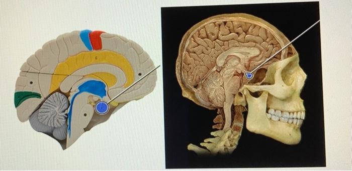
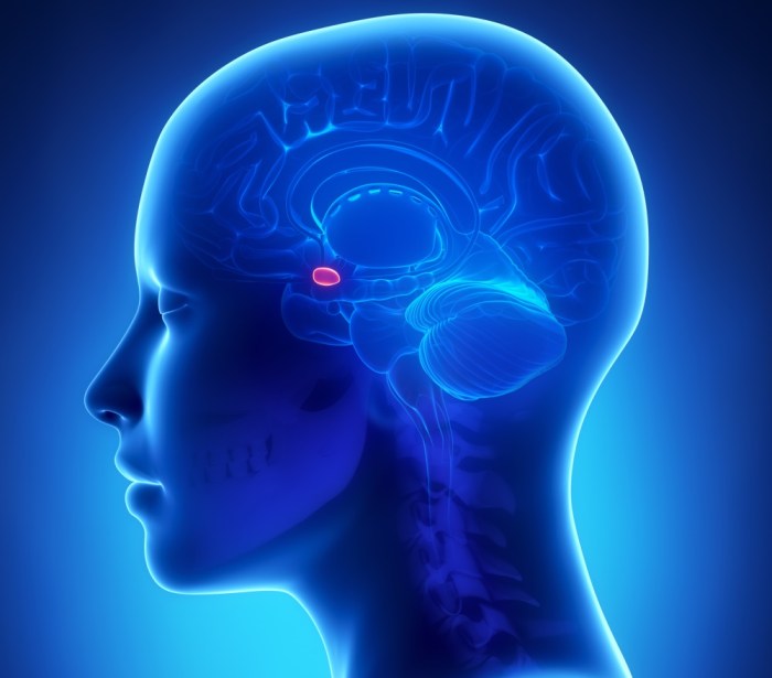
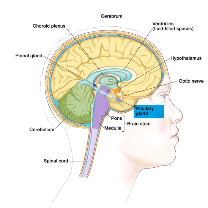
Which structure is highlighted pituitary gland – Which structure is highlighted: the pituitary gland? This question embarks us on an intriguing journey into the depths of human anatomy, revealing the intricate details and profound significance of this remarkable endocrine organ.
Nestled within the sella turcica of the sphenoid bone, the pituitary gland, despite its diminutive size, plays a pivotal role in regulating a myriad of bodily functions, making it a captivating subject of scientific exploration.
Anatomical Structure

The pituitary gland, also known as the hypophysis, is a small, pea-sized endocrine gland located at the base of the brain within the sella turcica, a bony cavity of the sphenoid bone.
The pituitary gland is roughly ovoid in shape, with an average size of 10-15 mm in length, 10-15 mm in width, and 5-8 mm in height. It is oriented in the midline of the body, just inferior to the hypothalamus and connected to it by the pituitary stalk.
Surrounding the pituitary gland are several important structures, including the optic chiasm, the cavernous sinuses, the internal carotid arteries, and the sphenoid sinus.
Internal Organization

Lobes of the Pituitary Gland
The pituitary gland is divided into two distinct lobes: the anterior lobe and the posterior lobe.
- Anterior lobe (adenohypophysis):The larger of the two lobes, the anterior lobe is composed of glandular tissue and is responsible for producing and secreting hormones that regulate various bodily processes, including growth, reproduction, and metabolism.
- Posterior lobe (neurohypophysis):The smaller of the two lobes, the posterior lobe is composed of nervous tissue and serves as a storage and release site for hormones produced by the hypothalamus.
Histological Composition
The anterior lobe of the pituitary gland is composed of several types of cells, including:
- Somatotrophs:Secrete growth hormone (GH)
- Lactotrophs:Secrete prolactin (PRL)
- Corticotrophs:Secrete adrenocorticotropic hormone (ACTH)
- Thyrotrophs:Secrete thyroid-stimulating hormone (TSH)
- Gonadotrophs:Secrete follicle-stimulating hormone (FSH) and luteinizing hormone (LH)
The posterior lobe of the pituitary gland is composed of two main types of cells:
- Magnocellular neurons:Synthesize and release oxytocin and vasopressin
- Parvocellular neurons:Synthesize and release corticotropin-releasing hormone (CRH)
Vascularization and Innervation, Which structure is highlighted pituitary gland
The pituitary gland is supplied by two main arteries: the superior hypophyseal artery and the inferior hypophyseal artery. These arteries form a capillary network that supplies the gland with blood.
The pituitary gland is innervated by the autonomic nervous system, with sympathetic and parasympathetic fibers originating from the hypothalamus.
Functional Significance: Which Structure Is Highlighted Pituitary Gland
Endocrine Functions
The pituitary gland is a master endocrine gland that plays a crucial role in regulating various bodily processes, including:
- Growth and development:GH stimulates linear growth and development.
- Reproduction:FSH and LH regulate reproductive function in both males and females.
- Stress response:ACTH stimulates the release of cortisol from the adrenal glands, which helps the body respond to stress.
- Thyroid function:TSH stimulates the thyroid gland to produce thyroid hormones, which regulate metabolism.
- Water balance:Vasopressin regulates water reabsorption in the kidneys, maintaining fluid balance.
- Milk production:PRL stimulates milk production in lactating women.
Feedback Mechanisms
The secretion of pituitary hormones is regulated by feedback mechanisms involving the hypothalamus and target endocrine glands.
For example, when the thyroid hormone levels in the blood are low, the hypothalamus releases thyrotropin-releasing hormone (TRH), which stimulates the pituitary gland to secrete TSH. TSH then stimulates the thyroid gland to produce more thyroid hormones, which in turn inhibits the release of TRH and TSH, thus maintaining thyroid hormone levels within a narrow range.
Clinical Implications
Dysfunction of the pituitary gland can lead to a variety of clinical conditions, including:
- Hypopituitarism:Deficiency of one or more pituitary hormones
- Hyperpituitarism:Excess production of one or more pituitary hormones
- Pituitary tumors:Benign or malignant growths that can affect pituitary function
Imaging Techniques

MRI and CT Scans
Magnetic resonance imaging (MRI) and computed tomography (CT) scans are the primary imaging modalities used to visualize the pituitary gland.
- MRI:Provides detailed images of the pituitary gland and surrounding structures, allowing for the assessment of size, shape, and signal characteristics.
- CT scans:Provide high-resolution images of the bony structures surrounding the pituitary gland, such as the sella turcica, and can detect calcifications or bony erosion.
Specific Features on Imaging
On MRI, the normal pituitary gland typically appears as a well-defined, homogeneous structure with a T1-hypointense and T2-hyperintense signal.
CT scans can show the size and shape of the sella turcica, as well as any erosion or expansion of the bone.
Surgical Considerations
Surgical Approaches
Surgical approaches to the pituitary gland include:
- Transcranial approach:Involves opening the skull to access the pituitary gland from above.
- Transsphenoidal approach:Involves accessing the pituitary gland through the nasal cavity and sphenoid sinus.
Indications and Contraindications
Surgery may be indicated for pituitary tumors that are causing symptoms or for pituitary dysfunction that is not responsive to medical treatment.
Contraindications to pituitary gland surgery include:
- Severe medical conditions that make surgery too risky
- Tumors that are too large or invasive to be safely removed
Potential Complications
Potential complications of pituitary gland surgery include:
- Damage to the pituitary gland, leading to hypopituitarism
- Damage to surrounding structures, such as the optic chiasm or carotid arteries
- Cerebrospinal fluid leak
- Infection
FAQ
What is the primary function of the pituitary gland?
The pituitary gland serves as the master endocrine gland, secreting hormones that regulate various bodily processes, including growth, metabolism, and reproduction.
What imaging techniques are commonly used to visualize the pituitary gland?
Magnetic resonance imaging (MRI) and computed tomography (CT) scans are frequently employed to obtain detailed images of the pituitary gland, aiding in the diagnosis and monitoring of various conditions.
What are the potential complications associated with pituitary gland surgery?
Pituitary gland surgery carries potential risks, including damage to surrounding structures, bleeding, and infection. Careful preoperative planning and meticulous surgical techniques are crucial to minimize these risks.